
Figure 1. Increased Caspase activity in mouse ES cells upon induction of Differentiation.
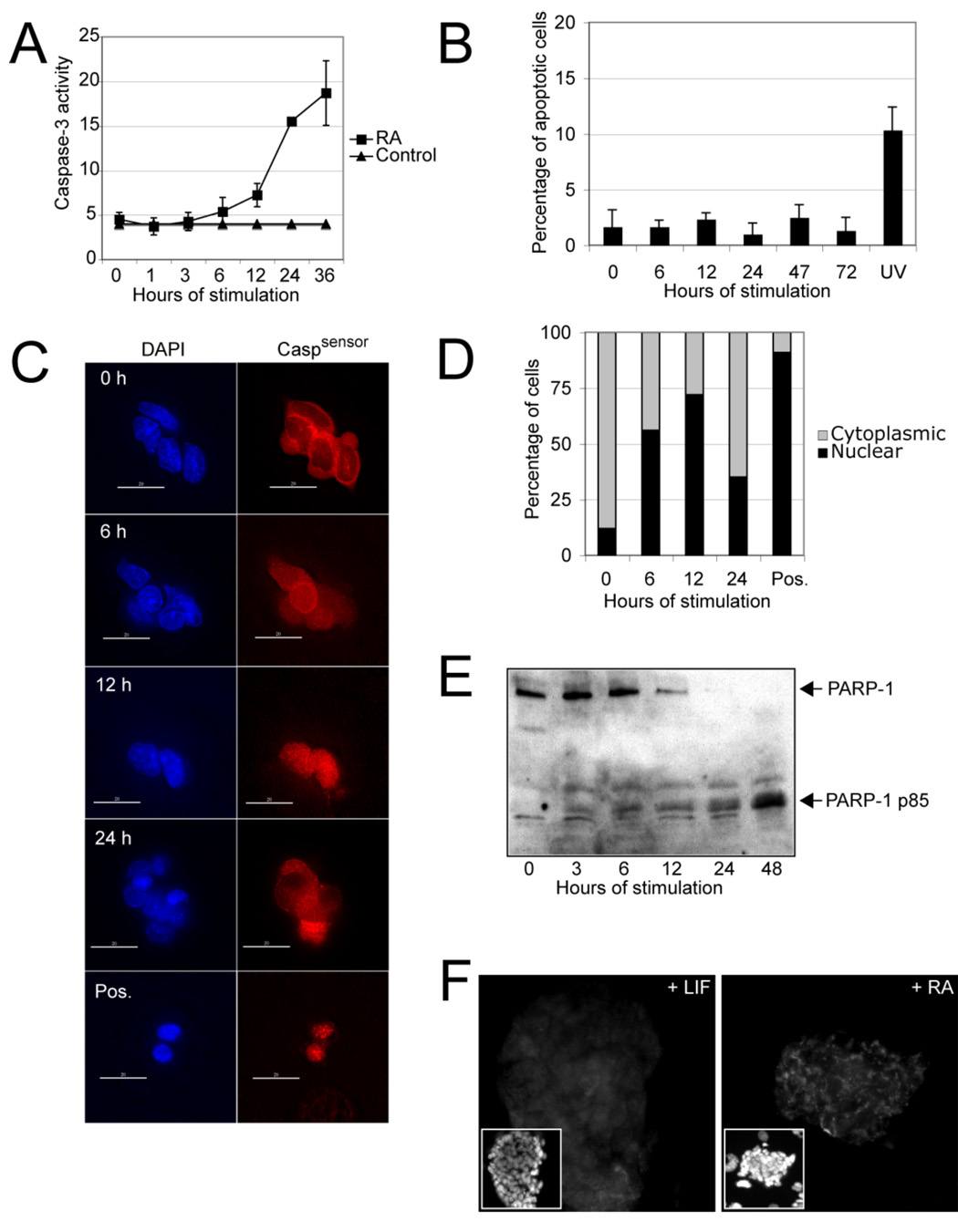
(A) The R1 ES cell line was stimulated with RA (1 µM) for the indicated times and Caspase activity was measured in an in vitro Caspase activity assay. The data are means (+/− SD) of triplicate experiments. (B) The same ES cell line was again exposed to RA for various times and the mean (+/− SD) percentage of cells undergoing programmed cell death was determined by counting apoptotic bodies . UV, ultraviolet light. (C) ES cells expressing the Caspase sensor (Caspsensor) were stimulated with RA (1 µM), fixed at the four indicated time points and stained with an antibody against a reporter protein (enhanced yellow fluorecent protein, EYFP). Mainly cytoplasmic staining indicates low Caspase acitivty, while mainly nuclear staining typically indicates increased Caspase activity. Immunofluorescence images (40X) were taken from representative fields. Although a shift of the signal from the cytoplasm to the nucleus is apparent at 12 hours, the cells did not show any signs of apoptosis. Cells treated with stausporine at 6 hours served as positive controls; scale bar = 10 µm. (D) Similar experimental setting as in panel C except that the mean percentage of cells with mainly cytoplasmic or nuclear staining was determined. (E) Western blot analysis of nuclear lysates isolated from mouse ES cells after stimulation with RA for the indicated times. The blotted membrane was probed with an antibody against PARP-1. The uncleaved form of PARP-1 is apparent at the earlier poststimulation times, with the cleaved form (85-kDa fragment) appearing after 24 hours, indicating PARP-1 cleavage. (F) Immunostaining of a mouse ES cell colony stimulated with RA or incubated with leukemia inhibitory factor (LIF) for 2 days. The antibody used specifically recognizes the 85 kDa form of PARP-1. Magnification, 40X.