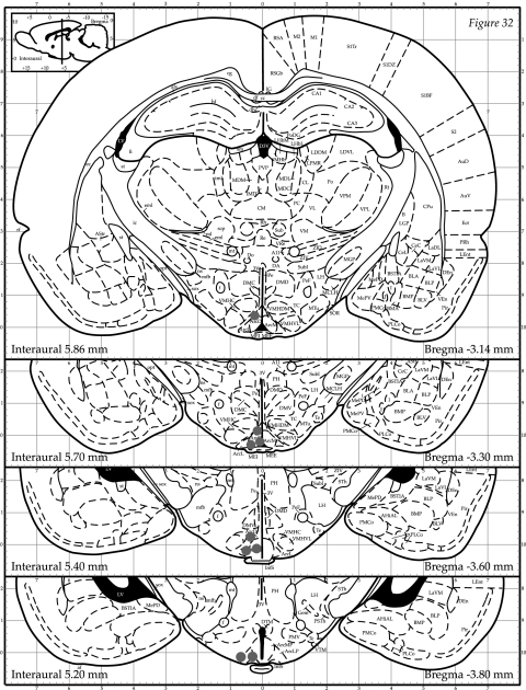
FIG. 5.
ARC and PVN cannula-placement sites. After completion of the hyperinsulinemic-euglycemic clamps, the animals were killed and toludine blue dye was injected into the cannula. The brain was then sectioned and placement was verified. Black dots represent correct cannula placement. Rats with cannulas not placed in the ARC and PVN were excluded from the analysis (n = 8).

