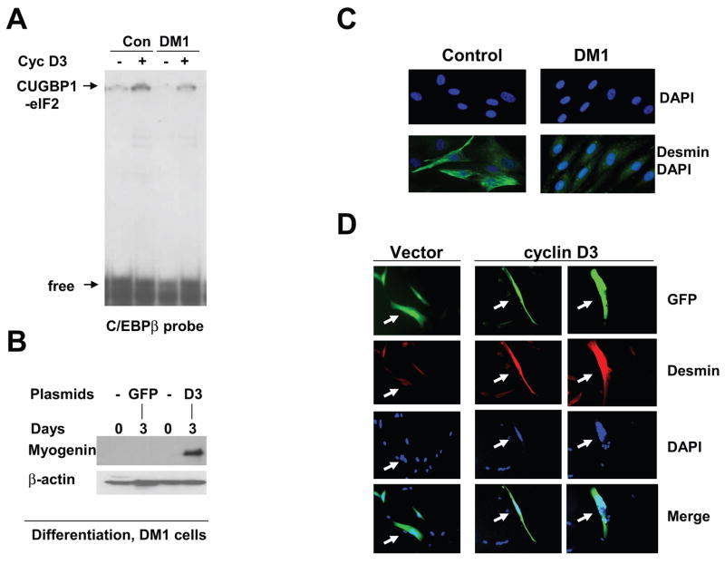
Figure 5. Increase of the levels of cyclin D3 in DM1 myoblasts corrects early steps of differentiation.
A. Ectopic expression of cyclin D3 in DM1 cells activates the CUGBP1-eIF2 pathway. Control and DM1 cells were transfected with pAdTrack-cyclin D3 plasmid, cytoplasmic extracts were isolated and used for EMSA with the C/EBPβ probe. Positions of the CUGBP1-eIF2 complex and free probe are shown by arrows. B. Ectopic expression of cyclin D3 increases protein levels of myogenin in DM1 differentiating cells. DM1 myoblasts transfected with GFP or cyclin D3/GFP were subjected to differentiation in fusion medium for three days. Myogenin levels were determined by Western blotting. The membrane was re-probed with antibodies to β-actin. C. DM1 cells do not increase expression of desmin at early step of differentiation. Immunofluorescent analysis with antibodies to desmin was performed using control and DM1 muscle cells differentiating for two days in fusion medium. Nuclei were stained with DAPI. C. Ectopic expression of cyclin D3 in DM1 cells increases expression of desmin during differentiation. DM1 myoblasts were transfected with pAdTrack-cyclin D3 plasmid expressing cyclin D3 and GFP from different promoters (green), differentiation was initiated by fusion media and cells were subjected to IF with antibodies to desmin (red). Nuclei were stained with DAPI. The picture shows representative analysis of 45 cells transfected with cyclin D3 and 41 cells transfected with an empty vector. Arrows show the transfected cells.

