
Figure 3. PRG-1 localizes to P granules during spermatogenesis.
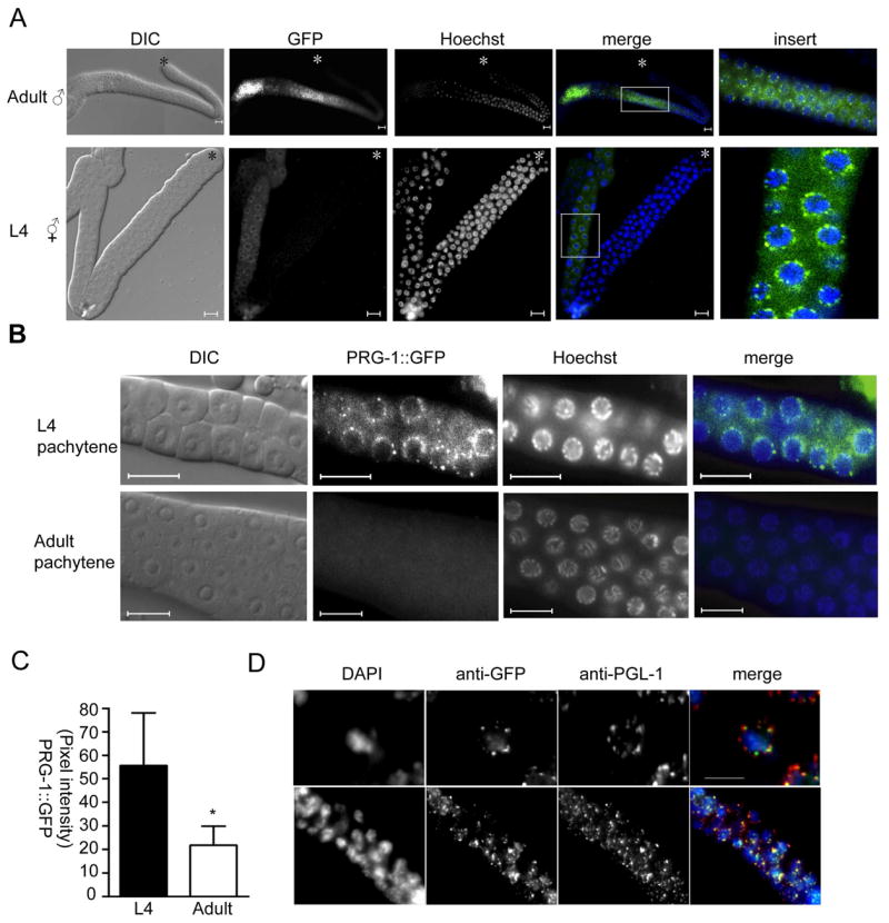
A) Live animals expressing the PRG-1:GFP transgene were exposed to Hoechst dye, and then gonads directly dissected and mounted for viewing. DIC and fluorescent images were collected. Scale bar: 10μm. B) The pachytene region of L4 and adult gonads from PRG-1::GFP transgenic animals are shown. Green, PRG-1::GFP; blue, Hoechst; scale bar, 10μm. C) PRG-1:GFP intensity was measured from equivalent exposures of pachytene germ cells from 10 animals each at L4 and adult stages. Error bar indicates standard deviation. * = Student’s t-test, P<0.01 D) L4 hermaphrodite gonads were stained with anti-GFP (green) and anti-PGL-1 (red). Upper panels: 1000X magnification; scale bar, 5 μm; lower panels: 400X magnification.