Fig. 3.
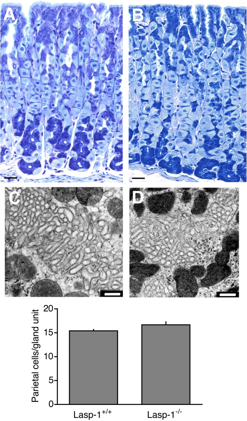
Histology, transmission electron microscopy (TEM) analyses, and parietal cell counts from gastric mucosal sections of 3 to 12-mo-old Lasp-1−/− and Lasp-1+/+ mice (A and B). Representative images of toluidine blue-stained gastric mucosal sections from Lasp-1−/− (A) and Lasp-1+/+ (B) male littermates (12 mo of age). Bars = 50 μm. C and D: representative TEM images showing intracellular canaliculi of parietal cells in gastric mucosae of histamine-stimulated mice. Three-mo-old Lasp-1+/+ (C) and Lasp-1−/− (D) littermates were injected as described in Fig. 2. Stomachs were processed 45 min later. Only the canalicular region, which is known to undergo changes in morphology in which F-actin-rich microvilli become elongated and cytoplasmic tubulovesicular numbers decrease, is shown. No differences in morphology were identified in other regions of the cells or in parietal cells from unstimulated controls. Bars = 500 nm. Graph: parietal cell numbers in gland units from Lasp-1+/+ and Lasp-1−/− male and female mice. Counts were performed on sections from 3-mo-old littermates: 1–3 sections/mouse, 5–25 glandular units counted/animal. No significant difference in parietal cell numbers was detected. N = 7 animals/group, P > 0.07.

