Fig. 5.
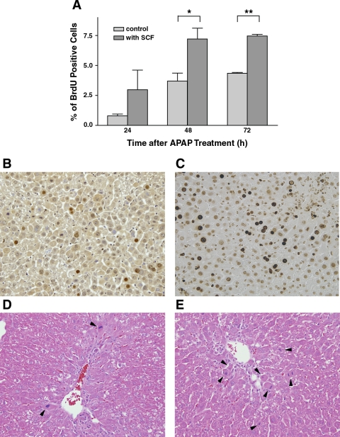
Effect of SCF on hepatocyte proliferation in vivo after acetaminophen treatment. C57BL/6 mice were treated with 375 mg/kg acetaminophen and SCF or PBS (vehicle control). Animals were killed at 24, 48, and 72 h postacetaminophen administration, and in vivo hepatocyte proliferation was estimated using a bromodeoxyuridine (BrdU) incorporation assay. A: animals treated with acetaminophen and SCF have a significantly increased rate of hepatocyte proliferation, compared with animals treated with acetaminophen and PBS, at 48 and 72 h (*P < 0.05 and **P < 0.01). B and C: representative histological sections after BrdU staining; B represents the liver from a mouse treated with 375 mg/kg acetaminophen with vehicle, and C represents liver from a mouse treated with 375 mg/kg of acetaminophen and SCF (48-h time point). Similarly, D and E are representative H&E staining of liver sections from mice treated with acetaminophen and PBS (D) or acetaminophen and SCF (E; both at 48-h time point). Arrowheads indicate the cells that are in mitosis; there are significantly more mitotic cells in the animal treated with SCF compared with the animal treated with PBS.

