Fig. 6.
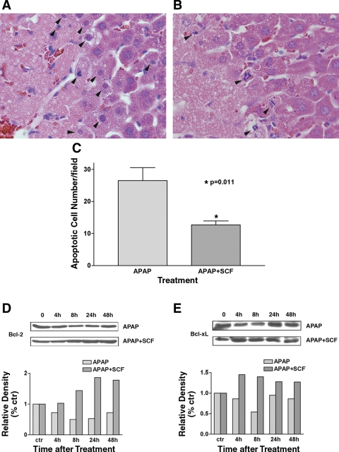
SCF inhibits acetaminophen-induced hepatocyte apoptosis. A: mice received 375 mg/kg acetaminophen and PBS (vehicle control), and liver tissue was obtained at 24 h. H&E staining demonstrates apoptotic hepatocytes between normal and necrotic hepatocytes (arrowheads indicate apoptotic cells). B: H&E staining of liver tissue obtained at 24 h from mice treated with 375 mg/kg acetaminophen and SCF; a decreased number of apoptotic hepatocytes are noted (arrowheads indicate apoptotic cells). C: apoptotic cells/high-power field in liver tissue 24 h after treatment with 375 mg/kg acetaminophen with (n = 6) and without (n = 6) SCF treatment. Ten fields were counted in each sample. Treatment with SCF results in a significant decrease in apoptotic cells. D and E: expression of Bcl-2 (D) and Bcl-xL (E) was examined with Western blotting. The gels represent 1 of 3 repeated experiments (all with similar results). The relative densities of Bcl-2 and Bcl-xL bands were normalized to those of GAPDH in the same samples.

