
Fig. 8.
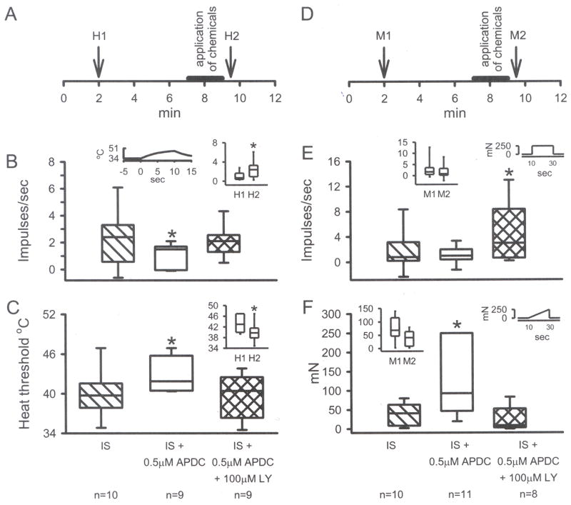
A) Standard in vitro paradigm with unit responses to heat recorded before (H1) and after (H2) drugs are applied for 2 min. IS increased the discharge rate of H2 compared to H1 (B inset) and decreased heat threshold to firing for H2 compared to H1 (C inset, *p<0.05, Wilcoxon test). Addition of 0.5 μM APDC blocked the IS-induced changes and 100 μM LY prevented the APDC-induced inhibition (B and C, p<0.05, *significantly different from all other groups, Kruskal-Wallis test). D) Standard paradigm with unit mechanical responses recorded before (M1) and after (M2) drugs are applied for 2 min. IS did not change the discharge rate (E, inset) or the threshold (F, inset) to mechanical stimulation. However, addition of APDC in the presence of IS increased the threshold (F, p<0.05, *different from all other groups, Kruskal-Wallis test) while addition of LY increased the discharge rate (E, p<0.05, * different from IS + APDC group only, Kruskal-Wallis test). Box plot values same as in Fig. 1.