Fig. 6.
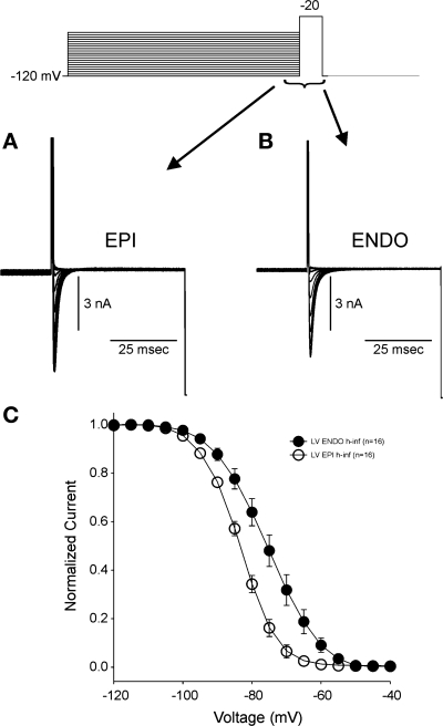
Representative steady-state inactivation recordings for Epi (A) and Endo (B) observed in response to the voltage-clamp protocol (top). C: steady-state inactivation relation for the two cell types. Peak currents were normalized to their respective maximum values and plotted against the conditioning potential. Epi cells showed a midinactivation potential that was significantly hyperpolarized compared with Endo cells.

