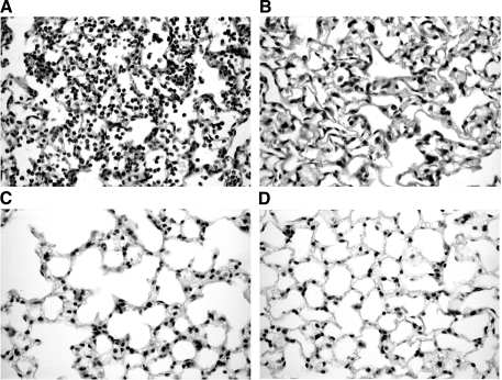
Fig. 3.
Representative light micrographs (×20) of hematoxylin and eosin-stained lung sections. A: 6 h after a mouse received IT IL-1β+TNF-α. The micrograph shows evidence of severe lung injury with an acute inflammatory response characterized by mononuclear inflammatory cell infiltrate and innumerable sloughed pneumocytes in the alveoli. The alveolar wall is edematous. Type II pneumocytes lining the alveoli are hypertrophic and hyperplastic. B: 6 h after a mouse received IT sterile saline (placebo). The micrograph shows empty alveolar spaces filled with air. The alveolar walls are lined with flattened epithelial cells. There are no inflammatory cells, no reactive type II pneumocytes, no fibrin, and no evidence of injury. C and D: 24 h after a mouse received either IT IL-1β+TNF-α (C) or IT sterile saline (D). Both micrographs show normal cellular morphology and no evidence of injury.

