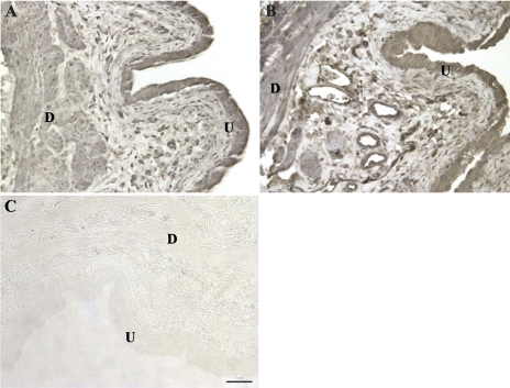
Fig. 5.
Immunohistochemical staining of NGF in the bladder wall 4 h after a single intravesical infusion of saline (A) or acrolein (B; acute cystitis). Specific staining of NGF was observed in the urothelium (U) and detrusor smooth muscle (D). Sporadic positive staining was also observed in leukocytes in the submucosal region of the bladder between the urothelium and detrusor. In the absence of specific antibody, no positive staining was observed (C). Staining for NGF was similar in all sections from control rats and rats with cystitis (acute and subacute). Original magnification ×20. Scale bar, 50 μm.

