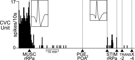
Fig. 6.
Inhibition of neurons in the rRPa eliminates spontaneous tail CVC neuronal activity and prevents tail CVC neuron activation by PGE2 in the POA. Spike frequency record of a tail CVC neuron illustrates elimination of spontaneous CVC neuronal activity following MUSC into rRPa (arrowhead). Left inset: average of 30 spontaneous CVC neuron action potentials prior to MUSC in rRPa (trace duration, 5.5 ms; peak amplitude, 30 μv). Subsequent PGE2 into the POA (arrowhead) failed to evoke tail CVC neuronal activity. Single electrical stimuli (stim rRPa, between arrowheads) in the rRPa evoked CVC neuron action potentials (right inset: average of 30 stimulus-evoked CVC neuron action potentials, trace duration: 5.5 ms, peak amplitude, 23 μv), indicating that the CVC neuronal recording was maintained. Transections (TRANS X) at bregma −2 mm and −4 mm did not evoke tail CVC neuron activity after MUSC into the rRPa.

