
FIGURE 7.
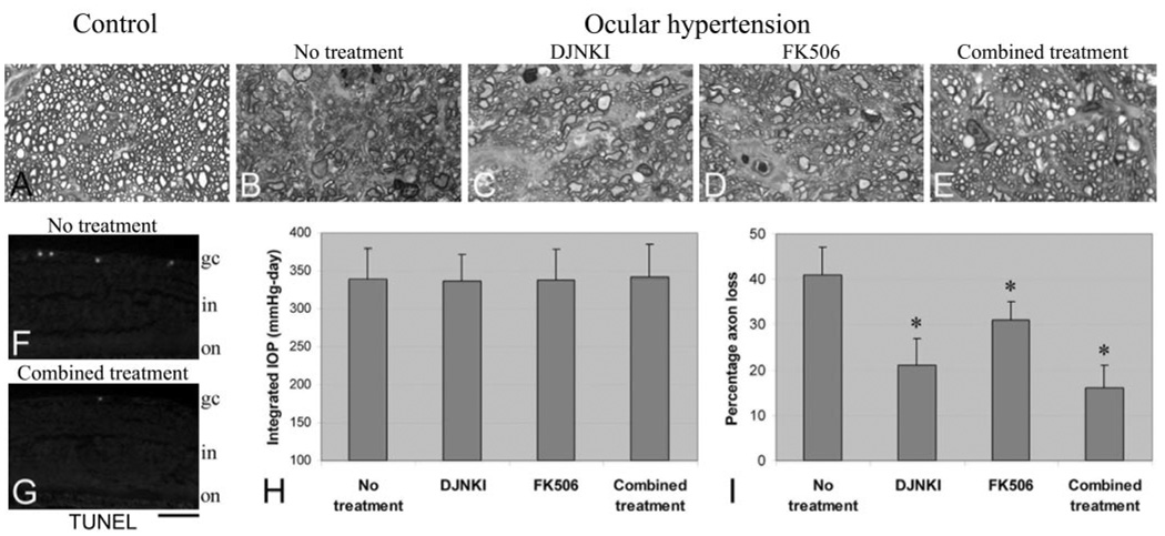
Optic nerve injury in ocular hypertensive eyes and protection by DJNKI and FK506 treatments. (A–E) Optic nerve cross-sections. Compared with those from normotensive control eyes (A), optic nerve cross-sections obtained from untreated ocular hypertensive eyes exhibited widespread degenerative changes characterized by axon loss, disorganized axon morphology, and myelin debris (B). However, the overall structure of the optic nerve was well preserved in treated ocular hypertensive rats (C–E). (F, G) Parallel to optic nerve findings, a decrease was also detectable in TUNEL-positive RGCs in the retinas of treated ocular hypertensive animals. Optic nerve axons were counted to quantitatively determine the treatment effect on neuronal damage. Axon counts revealed that although the treatments did not affect IOP in ocular hypertensive animals (H), they did result in a significant decrease in axon loss (I). Combined treatment with DJNKI and FK506 resulted in the greatest neuroprotective effect (combined treatment vs. no treatment; P = 0.0001). The neuroprotective effect of the combined treatment was found to be significantly greater than FK506 treatment applied alone (P = 0.001); however, the greater neuroprotective effect of the combined treatment was not statistically significant compared with DJNKI treatment applied alone (P = 0.1). gc, ganglion cell; in, inner nuclear; on, outer nuclear. *P < 0.05; statistically significant. Scale bar, 200 µm.