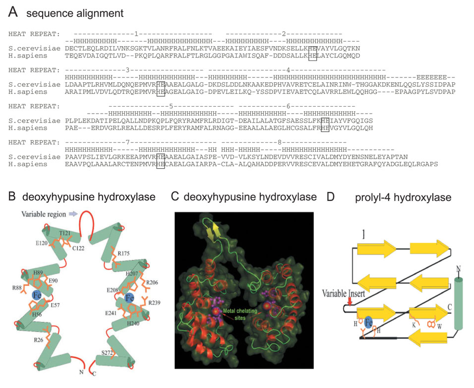
Fig. 5. Sequence and predicted structure of DOHH.
A: Sequence alignment of S. cerevisiae and human DOHH protein. A more complete comparison of DOHH sequences is given in Ref. 23. The conserved HE (HisGlu) pairs are boxed. Schematic diagram (B) and 3D model (C) of DOHH in comparison with prolyl 4-hydroxylase (D) Modified from Ref. 23.

