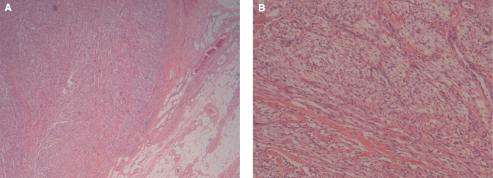
Figure 2.
Histological examination at low magnification demonstrating the clear boundary of the mass with the bladder wall (A) (hematoxylin–eosin stain, original magnification 100 ×). The tumour extends through the entire thickness of the bladder wall and focally is seen abutting the perivesical adipose tissue. High magnification of the mass demonstrating the eosinophilic spindled cells arranged in loosely cohesive fascicles (B) (hematoxylin–eosin stain, original magnification 200 ×).

