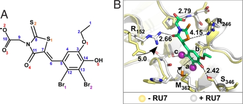
Fig. 3.
Structure of a small-molecule inhibitor bound to the β-clamp. (A) The RU7 compound. (B) Distances between RU7 and the β-clamp. Side-chain movements upon binding RU7 are indicated. Yellow and white are residues of β in the absence or presence of RU7, respectively. Distances between RU7 and protein residues are marked in black; the distances 3.73, 3.41, and 3.03 Å are marked a, b, and c, respectively.

