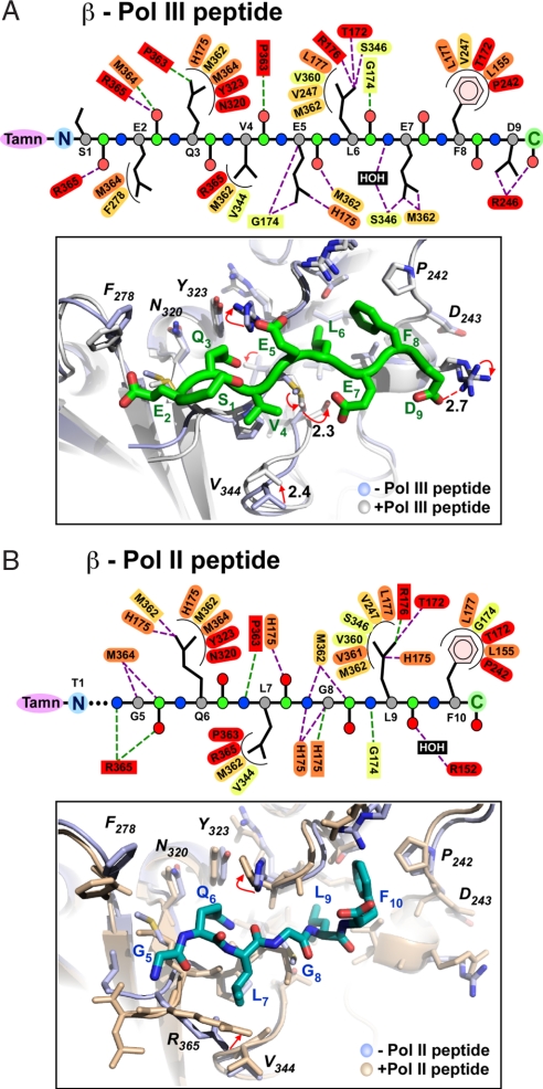
Fig. 4.
Structures of Pol II and Pol III peptides bound to the β clamp. (A Upper) Main-chain atoms of the Pol III peptide are shown as circles (C, green; Cα, gray; N, blue; and O, red). β-Clamp residues (ovals, side-chain interactions; rectangles, main chain) are colored according to conservation as described in the legend to Fig. 5. Hydrophilic and hydrophobic interactions are shown as straight purple and curved black lines, respectively. (Lower) Pol III 9-mer peptide (green) bound to β. Peptide-free β (light blue) is superimposed onto peptide-bound β (white). (B Upper) Pol II peptide atoms and β side chains are color coded as in A. (Lower) Pol II 10-mer peptide (blue) bound to β. Peptide-free β (light blue) is superimposed onto the peptide-bound β (wheat).

