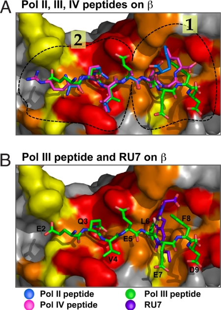
Fig. 5.
Superposition of Pol II, III, and IV peptides and RU7 compound bound to β. (A) Superposition of the Pol III peptide (green), Pol II peptide (blue), and Pol IV peptide (purple; PDB ID code 1OK7) (10). (B) Superposition of Pol III peptide (green) and the RU7 compound (orange). The surface of the β-clamp is colored white, and the protein-binding pocket of β is colored according to sequence conservation of an alignment of 42 bacterial subunits; the color scale proceeds from red (90% conservation) to yellow (50% conservation). Circled regions in the peptide-binding pocket indicate subsites 1 and 2. Figures were prepared by using Pymol (27).

