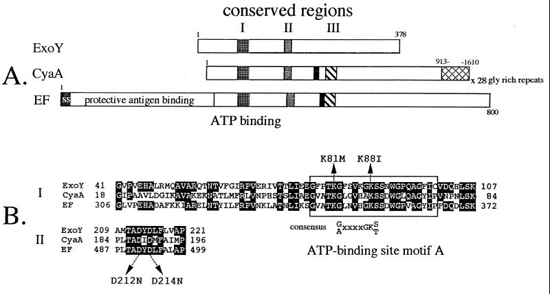
Figure 1.
(A) Schematic representation of the ExoY, CyaA, and EF adenylate cyclases. The positions of conserved regions I, II, and III are shown. Solid boxes represent the calmodulin-binding domains of CyaA and EF. The glycine-rich repeats of CyaA (hatched), signal sequence (ss), and protective antigen-binding site of EF are labeled. (B) pileup alignment of conserved regions I and II. Residues within CyaA and EF that are homologous to ExoY are shaded. The position of ATP-binding site motif A and its consensus sequence are shown. Residues of ExoY altered by site-directed mutagenesis are indicated by arrows.

