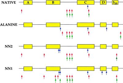
Figure 6.
Summary of proteolysis patterns for the native, nonnative, and alanine variants of MinLeu. The polypeptide chain is represented schematically, with the locations of each native helix highlighted in yellow. Proteolysis sites for each variant of MinLeu are indicated by rows of arrows beneath the polypeptide chain. Elastase, subtilisin, and proteinase K cuts are shown by blue, red, and green arrows, respectively (Top to Bottom).

