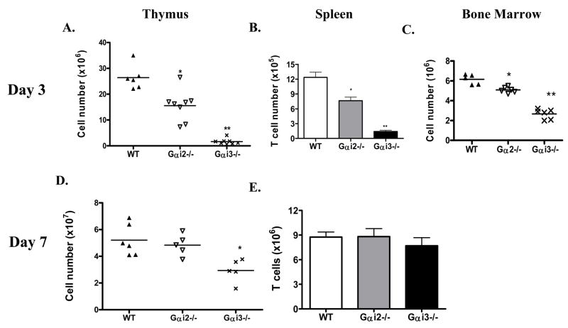
Fig. 1.
Abnormal thymocyte development in neonatal Gαi-deficient mice. Thymi (A), spleen (B) and bone marrow (C) were isolated from wild type (WT) and Gαi2−/− and Gαi3−/− mice at 3 days of age. Single cell suspensions were prepared and counted. T cell numbers in the spleen were obtained in the basis of percentages of CD3+ cells identified by flow cytometry. Each symbol in A, C and D relates to data obtained from a single animal and the data in B and E are the means ± standard deviation (SD) of 6 (WT), 8 (Gαi2−/−) and 7 (Gαi3−/−) mice. *, p<0.01 and **, p<0.001 in the presence or absence of Gαi2 or Gαi3.

