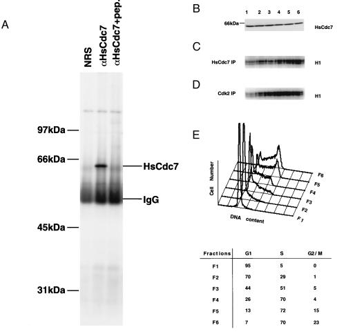
Figure 4.
The expression levels of HsCdc7 protein and its kinase activity during the cell cycle in 293 cells. (A) Lysates from 293 cells were immunoprecipitated with nonimmune rabbit serum (NRS), HsCdc7 antiserum or HsCdc7 antiserum that had been prebound to HsCdc7 C-terminal peptide. After washing, the immunoprecipitates were subjected to SDS/PAGE, transferred to Immobilon-P membrane and then blotted with HsCdc7 antiserum. (B) Subconfluent 293 cells were fractionated at specific stage of the cell cycle using centrifugal elutriation as described in Materials and Methods. Cell lysates from the indicated fractions (F1–F6) were subjected to SDS/PAGE, transferred to Immobilon-P membrane and then blotted with affinity-purified HsCdc7 antibodies. (C and D) The same cell lysates used in B were immunoprecipitated with HsCdc7 antibodies (C) or Cdk2 antibodies (D), and subjected to in vitro kinase reactions using histone H1 as substrate plus [γ-32P]ATP. The products of the kinase reactions were separated by SDS/PAGE prior to autoradiography. (E) 293 cells from each elutriated fraction as described in B were collected and analyzed for DNA content by flow cytometry. (Lower) The values represent the percentage of cells in the indicated phase(s) of the cell cycle in fractions (F1–F6), determined as described in Materials and Methods.

