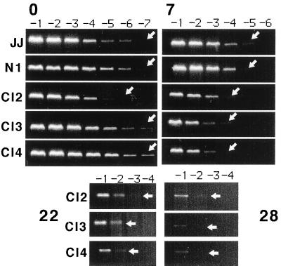
Figure 3.
Semiquantitative analysis of viral DNA in cell lines infected with cell-free HHV-6. DNA was extracted from Rep-transformed cell lines (Cl2, Cl3, Cl4), parental cells (JJ), and N1 control cell line at various time points after viral infection [either immediately postinfection (0), or at 7, 22, and 28 days postinfection]. Serial 10-fold dilutions of cellular DNA (starting at 0.1 μg) were analyzed by PCR, using primers specific for the viral U31 ORF. The arrows point to the highest dilution of template DNA yielding a positive signal upon PCR amplification.

