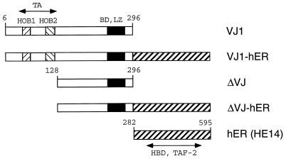
Figure 1.
Structure of v-Jun and v-Jun-ER fusion proteins. The basic DNA binding region (BD), the leucine zipper (LZ), and the homology boxes 1 and 2 (HOB1, HOB2) of Jun are shown as patterned boxes. Numbering of amino acid residues is according to c-Jun. The carboxyl-terminal half of the human ER HE14 (amino acids 282–595) comprising the hormone binding domain (HBD) and TAF-2 is depicted as a diagonally striped box. The v-Jun protein and the ER part are separated by a spacer consisting of the amino acids glycine-glycine-serine.

