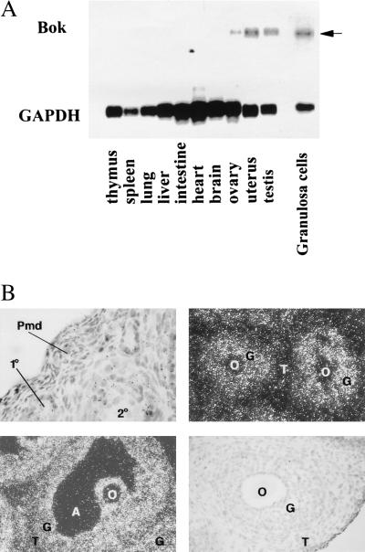
Figure 5.
Expression of Bok mRNA transcripts in rat tissues. (A) For Northern blot analysis, poly(A)+-selected RNA from different tissues of rats at 27 days of age or from isolated granulosa cells of estrogen-treated rats was hybridized with a 32P-labeled Bok cRNA probe. After washing, the blots were exposed to x-ray films at −70°C for five days. Subsequent hybridization with a glyceraldehyde-3-phosphate dehydrogenase cRNA probe was performed to estimate nucleic acid loading (8 h exposure). Specific Bok transcripts are indicated by an arrow. (B) In situ hybridization analysis of Bok expression in the ovary. Ovaries from immature equine chorionic gonadotropin-treated rats were probed with the antisense Bok cRNA. Upper, Left: hybridization signals in representative primary (1°) and secondary (2°) but not primordial (Pmd) follicles (magnification, ×100). Upper, Right (magnification, ×200) and Lower, Left (magnification, ×100): positive signals in granulosa cells of preantral follicles and an antral follicle, respectively. Lower, Right: no signal was found in a section hybridized with the sense Bok probe. O, oocyte; G, granulosa cells; T, theca cells; A, antrum.

