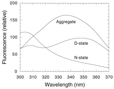
Figure 2.
Fluorescence of p53 core domain in 50 mM sodium phosphate, pH 7.2/5 mM DTT at 25°C on excitation at 280 nm. The native state (N) has a tyrosine emission maximum at 305 nm, which shifts to 310 nm on denaturation. There is a clear tryptophan emission maximum for the denatured state (D) at 350 nm. The tryptophan fluorescence of N is very weak. D and N have the same fluorescence at 321 nm. Aggregated states have a fluorescence maximum at 340 nm; shown is the emission of p53 core domain after being heated to 60°C and cooled to 25°C.

