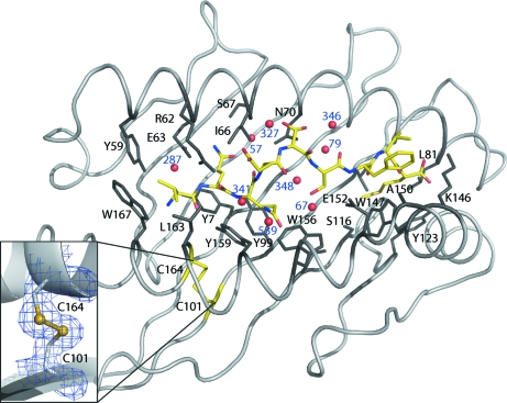
Figure 1.
Interactions in the HLA-B*1501 peptide-binding groove. The HLA-I complex is visualized from above, looking directly towards the floor of the peptide-binding groove. The peptide (VQQESSFVM) is depicted in yellow and the peptide N-terminal region is located to the left. Peptide-binding groove residues interacting with the peptide are coloured black and labelled. The insert shows the electron density (at 1σ, to a distance of 1 Å) of the characteristic disulfide bond in the α2 helix.

