
Fig. 2.
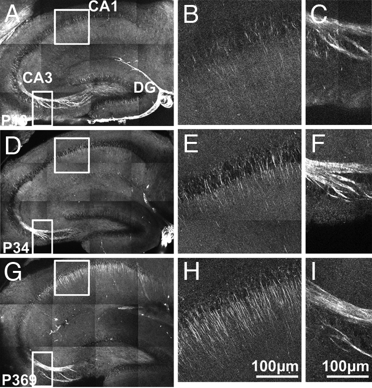
Polarized microtubule arrays in acute hippocampal slices of young, juvenile, and adult mice. (A) SHG in hippocampus of a postnatal day 13 (P13) mouse. The patchy background is caused by weak autofluorescence that is picked up by the SHG detector. (B and C) Magnified SHG views of the area CA1 and the mossy fiber from the boxed areas in A. (D–F) Same as A–C of a P34 mouse. (G–I) Same as A–C of a P369 mouse. Images are mosaics of 12 projections of optical z-sections at 15 μm apart with laser excitation at 774 nm that has linear polarization oriented along the apical dendrites.