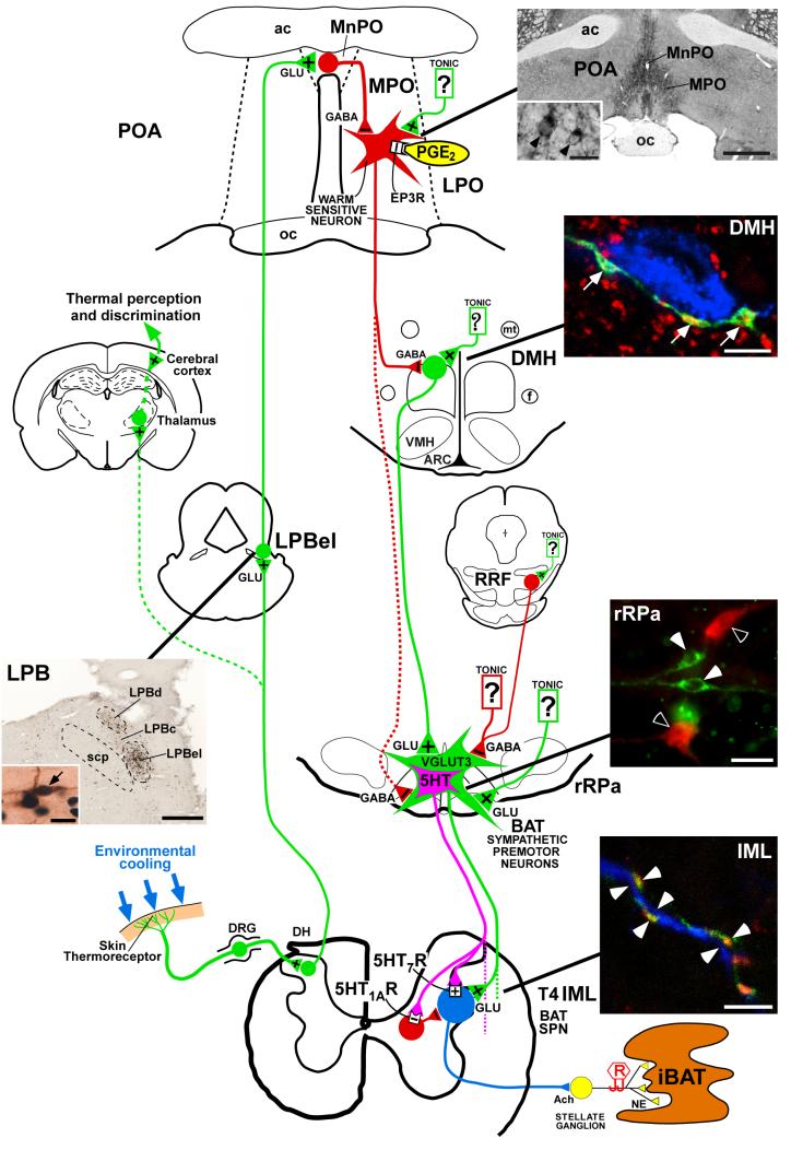
Figure 1. Schematic diagram summarizing the proposed model for the feedforward reflex circuit for cold-evoked brown adipose tissue thermogenesis.
Photomicrographs of the lateral parabrachial nucleus (LPB), the preoptic area (POA), the dorsomedial hypothalamus (DMH), the rostral raphe pallidus (rRPa) and spinal intermediolateral nucleus (IML) illustrate the anatomical substrates for key neurochemical and synaptic aspects of the proposed thermoregulatory circuit. In the LPB panel, (LPBel), central (LPBc) and dorsal (LPBd) subnuclei of the LPB contain neurons that are retrogradely labelled with a tracer (brown) from the median preoptic (MnPO) subregion of the POA. Retrogradely labelled neurons in the LPBel and LPBc, but not those in the LPBd, also express Fos (blue-black nuclei) following cold exposure of the animals (arrow, inset); scp, superior cerebellar peduncle; scale bars represent 0.5 mm and 15 μm (inset). Reproduced with permission from Nakamura & Morrison, 2008b). In the POA panel, immunohistochemistry for EP3 receptors (EP3R) shows the localization of these receptors in cell bodies (inset, arrowheads) and dendritic fibres of neurons that are distributed in the MnPO and medial preoptic (MPO) subregions of the POA; ac, anterior commissure; oc, optic chiasm; LPO, lateral preoptic area; PGE2, prostaglandin E2; GLU, glutamate; scale bars represent 1 mm and 20 μm (inset). Reproduced with permission from Nakamura et al. (1999). In the DMH panel, axon swellings of POA neurons (green) that are positive for a marker of GABAergic terminals (red) are closely apposed (arrows) to neurons that are retrogradely labelled with a tracer (blue) from the rRPa; ARC, arcuate nucleus; f, fornix; mt, mammillothalamic tract; VMH, ventromedial hypothalamic nucleus; RRF, retrorubral field; scale bar represents 5 μm. Reproduced with permission from Nakamura et al. (2005). The rRPa panel shows double immunofluorescence labelling for vesicular glutamate transporter 3 (VGLUT3)-positive (green, white arrowheads) and serotonin (5-HT)-positive neurons (red, open arrowheads); BAT, brown adipose tissue; scale bar represents 30 μm. The IML panel shows that axon swellings of rRPa neurons (green) that are positive for VGLUT3 (red) are closely associated (white arrowheads) with dendritic fibres positive (blue) for a marker of sympathetic preganglionic neurons (SPNs); Ach, acetylcholine; DH, dorsal horn; DRG, dorsal root ganglia; iBAT, interscapular BAT; NA, noradrenaline; R, recording electrode; scale bar represents 5 μm. Reproduced with permission from Nakamura et al. (2004a).

