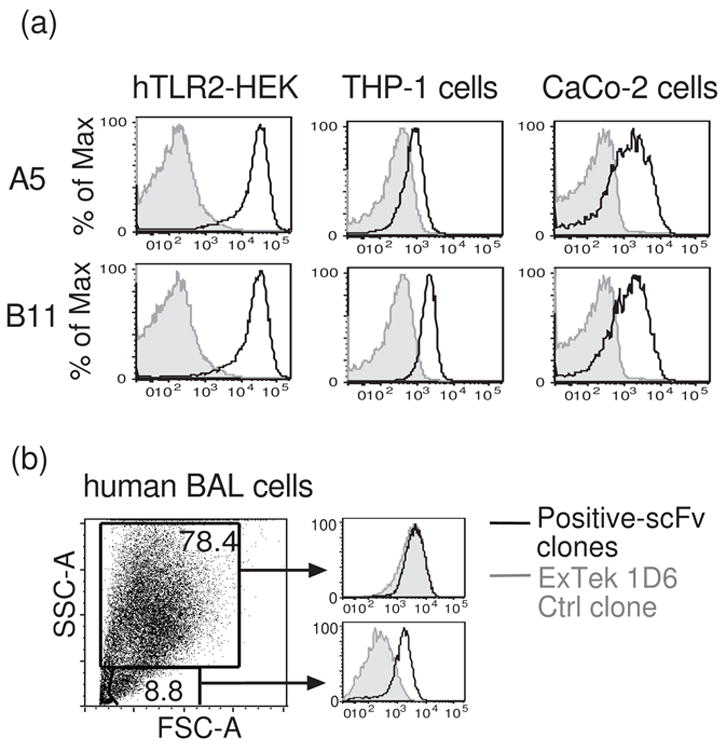
Figure 5.

Anti-hTLR2 scFvs specifically bind to endogenous hTLR2. (a) Flow cytometric profiles of the binding of two scFv clones to human THP-1 and CaCo-2 cells. The binding of the clones to hTLR2-HEK cells is shown for comparison. (b) Binding of an anti-hTLR2 scFv to human bronchial alveolar lavage (BAL) cells obtained during pulmonary inflammation. Only small SSC-low cells demonstrate specific staining for hTLR2. In each panel, signals obtained with anti-hTLR2 scFvs (black lines) were compared with signals obtained with an anti-ExTek negative control (gray shadows). Histograms are formatted as in figure 3a.
