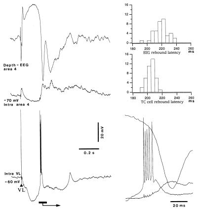
Figure 2.
Comparison between latencies of postinhibitory rebound in the VL neuron and field potential from the depth of area 4. Dual intracellular recordings from VL and area-4 neurons are shown, together with field potential from area 4. Recordings shown were made from cat under ketamine/xylazine anesthesia. (Left) VL stimulus elicits a postinhibitory rebound spike-burst in the VL cell followed by rebound depolarization in the area-4 neuron and depth-negative field potential in area 4. The rebound responses (marked by horizontal bar and arrow in Lower Left) are expanded in Lower Right; small deflections in intracellular recording from area 4 are caused by capacitive coupling from action potentials in the VL neuron. (Upper Right) Latency histograms of first action potential in rebound responses of the VL cell and peak negativity of field potential from area 4 (n = 40).

