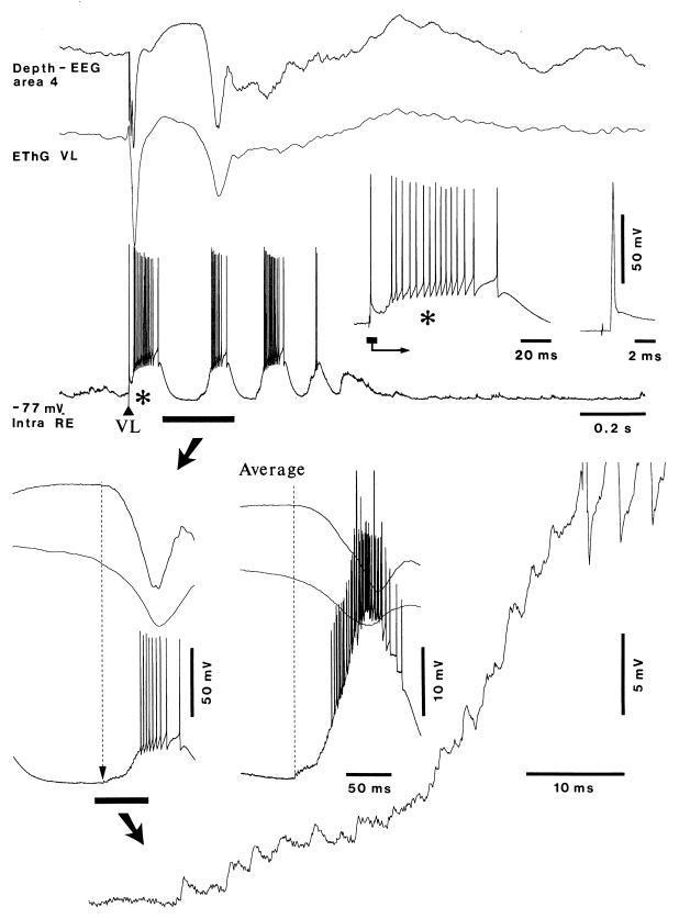
Figure 4.
Rebound EPSPs in the RE cell occur before rebound excitation in cortical area 4. Recordings shown were made from cat under ketamine/xylazine anesthesia. Intracellular recording of an RE cell from the rostrolateral sector of the RE nucleus is shown, together with field potentials from the VL nucleus and depth of area 4. (Upper) VL-evoked early response and spindle oscillation. (Upper Right) The early response in the RE neuron is expanded to show the initial antidromic discharge, followed by a high-frequency burst (indicated by an asterisk). (Lower Left) First postinhibitory excitation is expanded from the part marked by the horizontal bar and arrow in the Upper trace; the dotted line and arrow indicate the beginning of EPSPs in the RE neuron, well in advance of the onset of field negativity in area 4, but simultaneously with the developing rebound excitation in the field potential from the VL nucleus; below and to the right, RE-cell EPSPs are expanded further (as indicated by a horizontal bar and arrow). These EPSPs are triggered by rebound spike-bursts in TC cells (see text); EPSP-triggered average (n = 5) shows that EPSPs in the RE cell precede field negativity in area 4 (dotted line).

