
Figure 2. T cells form a dense infiltrate in the kidney.
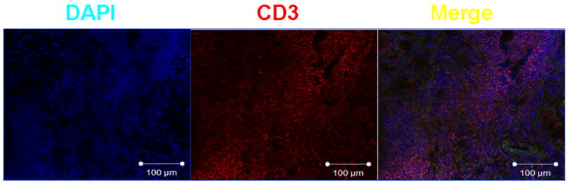
Frozen sections of the kidney biopsy were fixed with ice-cold acetone and incubated with mouse anti-human CD3 (1:100). After thorough washing, sections were incubated with goat anti-mouse IgG labeled with Texas Red (1:50). Finally, nuclei were stained by brief incubation with DAPI (4',6-diamidino-2-phenylindole; 0.5 μg/mL) (4). Slides were scanned in a Nikon Eclipse Ti confocal microscope; images were analyzed with EZ-C1 v. 3.6 software.