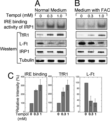
Fig. 2.
Tempol supplementation activated the IRE binding activity of IRP1, increased TfR1 levels, and decreased ferritin expression in mouse embryonic fibroblasts derived from IRP2−/− animals. (A) IRP2−/− embryonic fibroblasts were maintained in cultures supplemented with 0, 0.3, or 1.0 mM Tempol for 16 h. In cells that lacked IRP2, all IRE binding activity in the first row was attributable to IRP1 activation. Western blots revealed that TfR1 levels increased, and ferritin levels decreased in Tempol treated cells, whereas IRP1 and tubulin levels (loading control) did not significantly change. (B) IRP2−/− embryonic fibroblasts were maintained in cultures supplemented with 300 μM ferric ammonium citrate (FAC) and 0, 0.3, or 1.0 mM Tempol for 16 h. Increased IRE binding activity of IRP1 correlated with increased TfR1 levels and decreased ferritin levels. (C) IRE binding activities of IRP1 and TfR1 and L-Ft protein levels in the absence and presence of two different concentrations of Tempol without added FAC were quantified in comparison with the intensity of the control lanes, represented as 100%. Error bars represent the standard deviation calculated from the results of two different sets of experiments.

