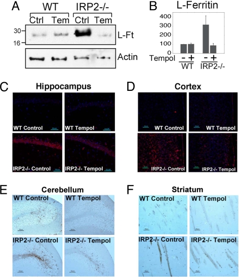
Fig. 4.
Increased IRE binding activity induced by Tempol treatment leads to decreased ferritin expression in brain and reduced ferric iron accumulation in white matter. (A) Ferritin levels detected in Western blot of cerebellar lysates of wild-type animals on a control (Ctrl) (lane 1) or Tempol (Temp) diet (lane 2) compared with IRP2−/− animals on a control (lane 3) or Tempol diet (lane 4) indicated that increased cerebellar ferritin levels of untreated IRP2−/− animals were markedly reduced by treatment with Tempol. (B) L-Ft protein levels in the cerebellum from IRP2−/− mice on a control diet and Tempol-supplemented wild-type and IRP2−/− mice were quantified relative to the intensity of the wild-type control mouse bands, represented as 100%. Error bars represent the standard deviation calculated from the results of three different sets of animals. (C) Ferritin immunohistochemistry performed on animals on control diets revealed marked increases in ferritin expression in hippocampal neurons in IRP2−/− animals (Lower Left), which decreased with Tempol treatment (Lower Right). (D) Tempol treatment diminished ferritin overexpression in the cortex of IRP2−/− mice. (E) Cerebellar folia from wild-type and IRP2−/− animals were stained with Perls' DAB stain for detection of ferric iron. Ferric-iron staining increased in the white matter of IRP2−/− animals on a control diet compared with wild-type controls, but decreased in IRP2−/− animals on the Tempol diet. (F) Ferric-iron staining was also increased in the striatum of IRP2−/− animals but diminished in IRP2−/− animals on Tempol supplementation.

