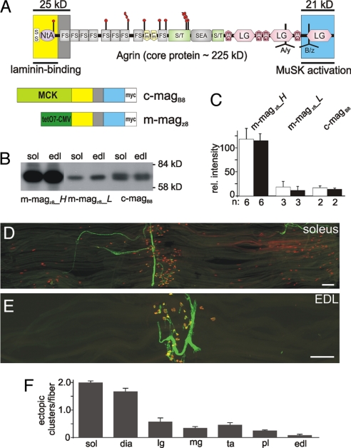
Fig. 1.
Transgenic expression of a miniaturized form of neural agrin induces the formation of nonsynaptic AChR clusters. (A) (Upper) Schematic scheme of the protein domains of agrin and the localization of the alternative mRNA splice sites A/y and B/z (modified from 12). (Lower) Schematic representation of miniagrin constructs used. Promoters that drive expression in skeletal muscle are shown in green and the domain encoded by the miniagrins in the colors using in the schematic scheme (Upper). Constructs encode a myc-tag (white) for detection. (B) Western blot analysis of muscle extracts of 6-week-old mice from different transgenic lines using anti-myc antibodies. Equal amount of protein was loaded into each lane and controlled by Ponceau S staining of the blot (not shown). (C) Quantification of the signals observed in Western blots (soleus: open bars; EDL: filled bars). Bars represent mean ± SD; n indicates number of samples. (D and E) Single fiber layer bundles of soleus (D) or EDL (E) muscles isolated from 6-week-old c-magB8 transgenic mice stained for AChRs (red) and motor nerves (green). (F) Quantification of the number of ectopic AChR clusters per muscle fiber. Numbers represent mean ± SEM; n = 3 mice. In each mouse between 87 and 424 muscle fibers were examined. sol: soleus; dia: diaphragm; lg: lateral gastrocnemius; mg: medial gastrocnemius; ta: tibialis anterior; pl: plantaris; edl: extensor digitorum longus. (Scale bars: 250 μm.)

