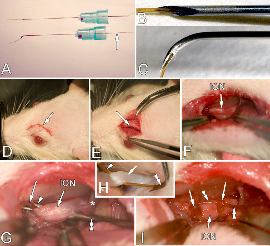
Figure 1.

A. A dental needle threaded with chromic gut suture (top neele) and with tip bent (bottom needle). The arrow indicates the needle extension found on dental needles that simplifies threading. B, C. High magnification of the threaded tip before and after bending, respectively. D. Location and size of the initial incision (arrow) E. Retraction of the skin to expose the edge of the frontal bone (arrow). F. Retraction of the orbital contents to show deep location of the ION (arrow). G. High magnification of the placement of the ligature. The syringe needle (double arrowheads) has been slid under the ION and the needle orifice (arrowhead) and protruding gut suture (arrow) is visible. The component of the opthalmic nerve (*) is also seen. H. This image is from a perfused rat with the orbit contents removed to more clearly show the end of the gut (arrowhead) that is held by the forceps (not seen) and the direction in which the needle (double arrowhead) is withdrawn to leave the gut suture in place. I. The ION with two ligatures (arrows) in place. The double arrowheads indicate the ends of the ligature remaining after tying which will be cut off before the skin is sutured closed. NOTE: In G and H the nerve is pulled up more than normal to show the procedure more clearly.
