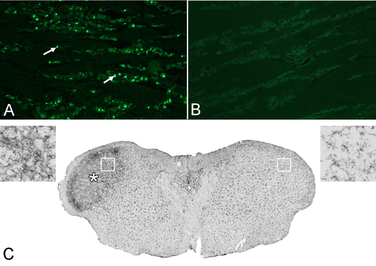
Figure 3.

Section from the trigeminal ganglion ipsilateral (A) and contralateral (B) to the CCI immunostained for the cell injury marker ATF3. CCI produces a significant increase in ATF3 immunopositive cells (e.g. arrows) in the ganglion on the injured side (labeled neurons/4×106 µm2; ipsi, = 144.16; contra, 2.58; p<0.001, n=4). A coronal section (C) of the caudal brainstem at the level of nucleus caudalis (5.0 mm posterior to interaural 0). Five days after CCI there is a significant increase in OX-42 immunostaining (*) on the side ipsilateral to the CCI (left. Pixel intensity, ratio ipsi/contra: 3.14; P < 0.001, n=4). The insets at top left and top right show higher magnifications of the areas indicated by the boxes.
