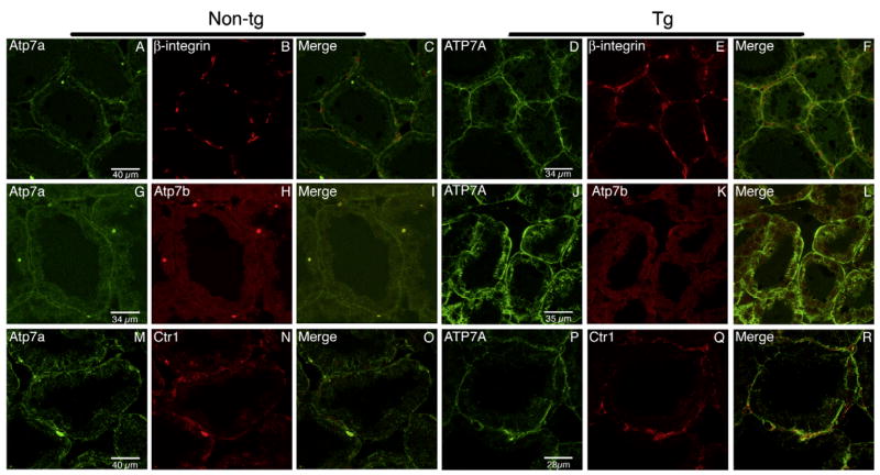
Fig. 3.
Immunofluorescence analysis of mammary gland tissue. Tissue sections were processed as described in Materials and methods and proteins were detected by indirect immunofluorescence with R17-BX (green) and β-integrin (red) (top panels), R17-BX (green) and anti-Atp7b (red) (middle panels), and R17-BX (green) and anti-hCTR1 antibodies (red) (bottom panels) using a Leica confocal microscope.

