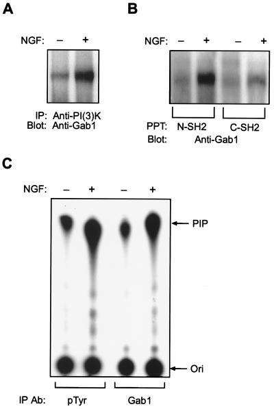
Figure 2.
Gab1 binds PI 3-kinase and mediates its enzymatic activation during NGF signaling. (A) PI 3-kinase interacts with Gab1. PC-12 cells were serum starved and mock stimulated or stimulated with NGF as indicated. Cell lysates were used for immunoprecipitation with a polyclonal antibody against PI 3-kinase (Upstate Biotechnology) and the Western blot was incubated with anti-Gab1 antibody. (B) The SH2 domains of p85 bind Gab1. Lysates from mock stimulated (−) or NGF stimulated (+) PC-12 cells were used for precipitations with glutathione S-transferase fusion proteins containing either the N-terminal SH2 domain (N-SH2) or the C-terminal SH2 domain (C-SH2) from the p85 subunit of PI 3-kinase. The blot was incubated with anti-Gab1 antibody. (C) Gab1 mediates PI 3-kinase activation during NGF signaling. PI 3-kinase assays were performed on anti-Gab1 and antiphosphotyrosine immunoprecipitates from mock stimulated or NGF stimulated PC-12 cell lysates. PIP, position of phosphotidyinositol-3 phosphate; Ori, origin. Results shown are representative of five independent experiments.

