Figure 4.
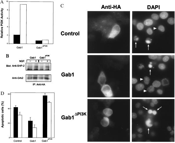
A Gab1 mutant lacking PI 3-kinase binding sites induces apoptosis in PC-12 cells. The Gab1 or Gab1ΔPI3K cDNAs were cloned into a pcDNA vector containing the HA epitope. These constructs or empty plasmid (Control) were transiently transfected into PC-12 cells (28). (A) Gab1ΔPI3K does not activate PI 3-kinase. PI 3-kinase assays were performed on the pellets from anti-HA immunoprecipitations and quantitated. Values were corrected for background and normalized for HA expression. Relative PI 3-kinase (PI3K) activity is shown. Solid bars, mock stimulated cells; open bars, cells treated with NGF. Results shown are representative of three different experiments. (B) Gab1ΔPI3K binds Grb2 and SHP-2 similar to wild-type Gab1. Western blots containing anti-HA immunoprecipitations were incubated with either anti-Grb2 or anti-SHP-2. (C) Gab1ΔPI3K induces apoptosis. Immunofluorescence microscopy was performed to detect cells expressing the various constructs (Anti-HA) while the nuclei were visualized using 4′-6-diamidino-2-phenylindole staining. Cells undergoing apoptosis are indicated by arrows and nuclei corresponding to the HA positive cells are designated by arrowheads, except in the case of Gab1ΔPI3K where they are co-incident. (D) Quantitation of the cells undergoing apoptosis. Cells were transiently transfected with these plasmids and then grown in serum containing media followed by growth in serum-free media or media supplemented with NGF for 5 h. The percentage of HA positive cells undergoing apoptosis is shown. The incidence of cell death observed with the control plasmid is similar to that reported by the manufacturer. Results are the average of six independent experiments. Solid bars, cells grown in serum free media; open bars, cells grown in media with NGF. Bars = SEM.

