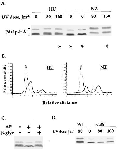
Figure 4.
Pds1p-HA is phosphorylated after the UV-irradiation of S phase-arrested cells. (A) Wild-type (OCF1522) cells were arrested in S phase with HU (0.1 M) or in mitosis with nocodazole (NZ, 15 μg/ml) and UV-irradiated at the indicated doses. Twenty minutes after irradiation, the cells were harvested and analyzed by Western blot analysis for Pds1p-HA. (B) Densitometry scans of the lanes indicated by asterisks in A: HU-treated cells, 0 and 160 J/m2; NZ-treated cells, 0 and 160 J/m2. Scans were done as described in Fig. 1B. The dashed and solid lines are of Pds1p-HA from nonirradiated and irradiated cells, respectively. (C) Wild-type (OCF1522) cells were arrested in S phase with HU (0.2 M) and UV-irradiated at 160 J/m2. The irradiated cells were harvested 20 min after irradiation and processed for phosphatase treatment. The phosphatase reaction were carried out in the presence (+) or absence (−) of 40 units of calf intestine alkaline phosphatase (AP) and 0.2 M β-glycerophosphate. (D) Wild-type (OCF1522) and rad9 (OCF1544) cells were arrested in S phase with HU (0.1 M) and UV-irradiated at the indicated UV doses as described in A.

