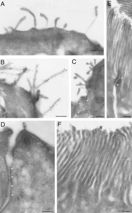
Figure 2.
Immunoelectron microscopy (26) of the 13A4 antigen (prominin) on the apical surface of neuroepithelial cells and the brush border of kidney tubules. Ultrathin cryosections (26) of paraformaldehyde-fixed tissue were stained with mAb 13A4 (0.5–1 mg/ml) followed by rabbit anti-rat IgG/IgM and 9-nm protein A-gold. Neuroepithelial cells of 9-day-old (A and C) and 10-day-old (B and D) mouse embryos and proximal tubule cells of adult mouse kidney (E and F) are shown. In neuroepithelial cells (A–D), immunoreactivity is confined to protrusions of the apical plasma membrane including microvilli-like structures. Junctional complexes are indicated by white arrowheads in C and D. In the kidney proximal tubule (E and F), immunoreactivity is restricted to the brush border where it appears to be concentrated at the tips of the microvilli. A tight junction is indicated by white arrowheads in E. (Bars = 0.5 μm.)

