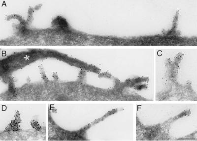
Figure 7.
Immunoelectron microscopy of prominin on the plasma membrane of transfected, paraformaldehyde-fixed CHO cells. Ultrathin cryosections were stained with mAb 13A4 followed by rabbit anti-rat IgG/IgM and 9-nm protein A-gold. The white asterisk in B indicates a filopodium extending from the cell shown in this panel. All panels are the same magnification. (Bar in F = 0.2 μm.)

