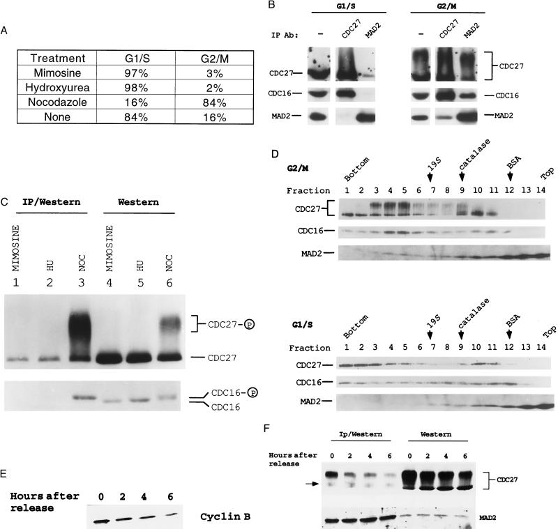
Figure 1.
MAD2 is associated with the cyclosome/APC in vivo. (A) The cell cycle profiles of HeLa cells treated with either 300 μM mimosine or 2 mM hydroxyurea (HU) for 12 hr, or with 100 nM NOC for 24 hr. HeLa cells were subjected to flow cytometry analysis as described (28). The cell cycle profile of untreated HeLa cells is also shown. (B) Coimmunoprecipitation of the cyclosome and MAD2 in HeLa cells. Extracts (1.5 mg) from HeLa cells arrested in G1/S with mimosine or G2/M with NOC were immunoprecipitated with either anti-MAD2 or anti-CDC27. The immunoprecipitates and HeLa extracts (75 μg per lane) were subjected to Western blot analysis as indicated. (C) MAD2 preferentially associates with the phosphorylated cyclosome in G2/M. Extracts (1.5 mg) from HeLa cells arrested in G1/S or G2/M with the indicated treatments were immunoprecipitated with anti-MAD2. The immunoprecipitates (lanes 1–3) and HeLa extracts (lanes 4–6, 40 μg per lane) were subjected to low-percentage SDS/PAGE to resolve the phosphorylated CDC16 from the unphosphorylated one. Anti-CDC16 reacted with two closely migrating bands in G2/M extracts, suggesting that a portion of CDC16 was phosphorylated (12) (lane 6, bottom row). (D) MAD2 and the phosphorylated cyclosome cosediment in glycerol gradients as a large complex. HeLa cells were arrested in G1/S with mimosine and in G2/M with NOC. Positions of the 19S regulatory complex of the 26S proteasome, catalase (232 kDa), and BSA (67 kDa) are shown. (E) Immunoblot analysis of the cyclin B levels after NOC release. HeLa cells were collected at the indicated time points after release from mitotic arrest. Equal amounts of extracts were then subjected to immunoblot analysis. (F) MAD2 dissociates from the cyclosome once the mitotic checkpoint becomes inactivated. Extracts were either directly subjected to Western blot analysis (Right, 60 μg per lane) or immunoprecipitated (1 mg extract) with the affinity-purified anti-MAD2 and the immunoprecipitates were then subjected to immunoblot analysis (Left). A background band in anti-MAD2 immunoprecipitates is indicated by an arrow. Note that anti-MAD2 antibodies primarily immunoprecipitated the phosphorylated forms of CDC27.

