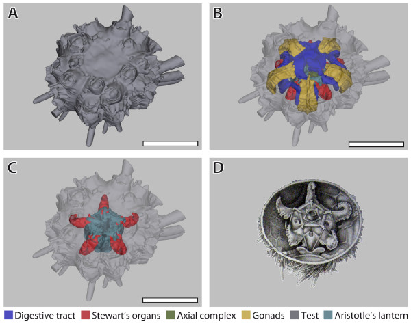
Figure 6.
Comparison of a digital 3D model with a traditional anatomical sketch. (A)-(C) Eucidaris metularia. Selected views taken from the interactive 3D model: (A) external view; (B) external view with transparent test, internal organs visible; (C) external view with transparent test and all internal organs removed except for Stewart's organs and Aristotle's lantern. (D) Cidaris cidaris (= Dorocidaris papillata). Image taken from [42] and modified. Stewart's organs constitute extensions of the peripharyngeal coelom. Scale bar: 1 cm. The colour legend specifies organ designation. The interactive 3D mode can be accessed by clicking onto Figure 6 in the 3D PDF version of this article: Additional file 7 (Adobe Reader Version 7.1 or higher required).

