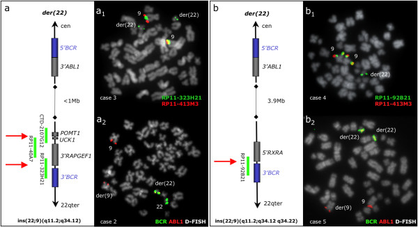
Figure 2.
BCR/ABL1 fusion at 22q11.2: "small" and "large" insertions with recurrent distal breakpoints. (a) Diagram showing the small size ins(22;9)(q11;q34.1q34.1) seen in 3 patients (no 1–3). The BAC clones covering the distal breakpoint region are presented with green lines. The ABL1 breakpoint marks the proximal boundary of the insertion (< 1 Mb) while the distal breakpoint (shown by red arrows) falls within a 280 Kb breakpoint cluster housing the UCK1, POMT1 and RAPGEF1 genes. (a1) A representative metaphase cell in patient no 3 with co-hybridization of FISH probes RP11-323H21 and RP11-413M3, showing a split signal from RP11-323H21 (green signals at both chromosome 9 homologues and masked Ph) and duplication of the masked Ph (green signals on 2 masked Ph). (a2) BCR/ABL1 D-FISH (Vysis) in patient no 2, showing the absence of green signal at der(9). (b) Diagram showing the large size ins(22;9)(q11.2;q34.1q34.2) seen in patient no 4 and CML-T1. The ABL1 breakpoint marks the proximal boundary of the 3.9 Mb insertion, while the distal breakpoint lies within the clone RP11-92B21 (red arrow). (b1) A representative metaphase cell in patient no 4 with co-hybridization of FISH probes RP11-92B21 and RP11-413M3, showing a split signal from RP11-92B21 (green signal at both chromosome 9 homologues and masked Ph). (b2) BCR/ABL1 D-FISH (Vysis) in CML-T1, showing absence of green signal at der(9) and duplication of the masked Ph (two fusion signals).

