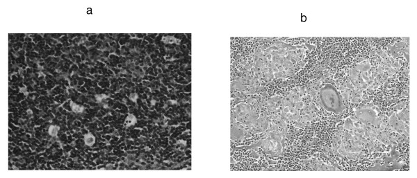
Figure 3.
a. Mass from the anterior mediastinum confirming thymoma B1 WHO type, lymphocyte rich predominantly cortical. (H&E stain, high magnification). b. Biopsy from the lymph nodes showing multiple non-caseating granulomas with multinucleated giant cells and histiocytes (H&E stain, low magnification).

