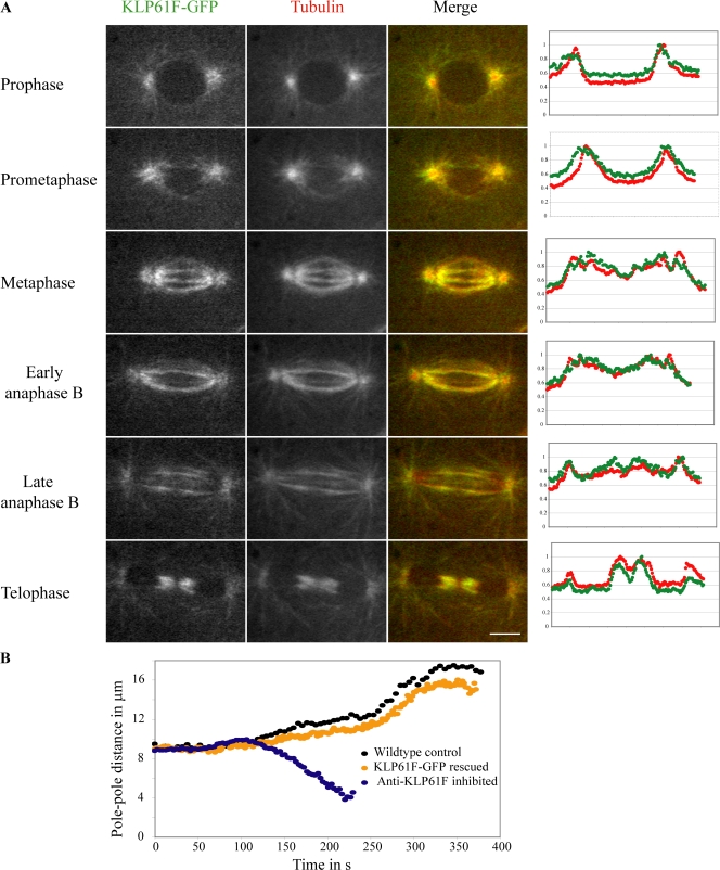
Figure 1.
Localization and function of KLP61F-GFP in D. melanogaster embryo mitosis. (A) Micrographs from a time-lapse video of a representative spindle showing KLP61F-GFP (left), rhodamine-tubulin (center), and double-label fluorescence (right) at various stages of mitosis. The plots (far right) are line scans extending pole to pole along an ipMT (10 pixels wide; ∼0.129 μm/pixel) for KLP61F (green) and tubulin (red). The y axis shows normalized fluorescence intensity. Bar, 5 μm. (B) Spindle pole dynamics in wild-type embryos, GFP-KLP61F rescued mutant embryos, and anti-KLP61F microinjected wild-type embryos showing how bipolar spindles collapse into monoasters after the loss of KLP61F function. Pole–pole separation dynamics are very similar in wild-type and rescued mutant embryos.

