Figure 3.
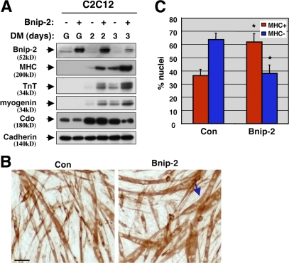
Overexpression of Bnip-2 enhances myogenic differentiation. (A) Lysates of C2C12 cells stably transfected with a control expression vector lacking a cDNA (−) or with an expression vector harboring a Bnip-2 cDNA (+) were Western blotted with antibodies to Bnip-2, the indicated muscle-specific proteins, or Cdo and, as a control, with pan-cadherin antibody. (B) Photomicrographs of C2C12/Bnip-2 and vector control cells that were cultured in DM, fixed, and stained with an antibody to MHC. The arrow indicates a C2C12/Bnip-2 cell myotube with many more nuclei than are seen with the control cells under these conditions. Bar, 0.1 mm. (C) Quantification of myotube formation. Values represent means of triplicate determinations ±1 SD. The experiment was repeated three times with similar results. Asterisks indicate difference from control, P < 0.01. A level of myotube formation by control cells (∼35% nuclei in MHC+ cells) was selected so as to permit visualization of enhanced differentiation by Bnip-2.

